
Ultrasound uses high-frequency sound waves to create images of your internal organs on a monitor. Ultrasound can help your doctor monitor your ovulation and diagnosis conditions such as pelvic masses and early pregnancy. Your doctor can also examine the thickness and pattern of your uterine lining.
Ultrasound is also used to monitor cycles during which you are taking fertility drugs to stimulate the follicles in your ovaries to produce multiple eggs. It is also used during in vitro fertilization (IVF) to help your doctor guide a needle into the follicle to retrieve eggs during vaginal oocyte retrieval (VOR), or to guide the catheter during embryo transfer.

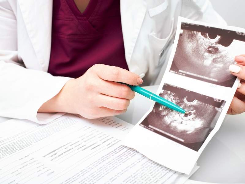
Ovary showing multiple follicles
How is an ultrasound performed?
Ultrasound uses high-frequency sound waves to create images of our reproductive organs on a monitor. Transvaginal ultrasound is performed with a scanning probe inserted into your vagina.
The probe is covered and a lubricant is used before it is inserted. The transvaginal ultrasound is inserted a short distance from your reproductive organs and produces very sharp, clear images on the monitoring screen. Even the smallest fibroids and certain other abnormalities can be detected. Ultrasound examinations are relatively painless and take only a few minutes to perform.
Questions & Answers

Trilaminar Uterine Lining
What does a transvaginal ultrasound feel like?
The scanning probe feels much like a tampon as it is inserted into your vagina. Once it’s completely inserted you may feel some slight pressure. If you are allergic to latex, be sure to tell your doctor, nurse, and technician before the test.
Does my bladder have to be full or empty during the test?
For a transvaginal ultrasound, you will empty your bladder just before the procedure.
When will I get the results of the test?
The results are ready as soon as the test is over. At RMA all ultrasound tests are performed by a doctor who will briefly review the findings and next steps in your treatment.